Clinically Relevant Anatomy
The patellofemoral joint makes part of the knee joint. The articular surfaces consist of the patella and the trochlear surface of the femoral condyles. The articular cartilage on the medial facet is thicker than on the lateral facet, with the lateral facet bigger than the medial. It has an anterior projection on the lateral femoral condyle, lateral to the patellar groove. This prevents lateral dislocation of the patella. The patellofemoral articulation depends on the function of the quadriceps as it increases the angle of pull of the patellar tendon, improving the mechanical advantage of the quadriceps in knee extension.
The suspension and movement of the patella is provided by passive and active stabilizers:
- Passive stabilizers: Tensor fascia lata, patellar ligament, knee capsule, patellofemoral ligament (medial and lateral), menisco patellar ligament (medial and lateral)
- Active: Quadriceps, patellar ligament, retinaculum
The medial patellofemoral ligament is the primary stabiliser (53-67%) against lateral displacement/dislocation of the patella. It is situated deep to the vastus lateralis muscle, ranging from the posterior aspect of the medial femoral condyle to the superomedial part of the patella, vastus medialis and quadriceps tendon.
Epidemiology/Etiology
Epidemiology
The incidence of acute primary patellar dislocations are 2-3%. Patellar dislocations are often associated with athletes, and is most common in females in the second decade of life. Redislocation rates after conservative management are estimated between 15 and 44%.
Aetiology
Primary patellar dislocation is defined as traumatic disruption of the previously uninjured medial peripatellar structures. It often results from a non-contact injury to the knee.
Predisposing factors include both morphological and functional patellofemoral disorders:
- Ligament laxity (can lead to atraumatic dislocations)
- Reduced osseous constraint form the lateral femoral condyle
- Imbalance between stronger lateral tissues (e.g. lateral retinaculum and vastus lateralis), which are able to overcome weaker medial structures, especially the medial patellofemoral ligament and the distal vastus medialis
- Biomechanical issues such as femoral and tibial rotation, and pes planus
- Patella alta
- Genu recurvatum
- Icreased Q-Angle
- Patellar hypermobility
Mechanism of Injury
- Non-contract: Twisting of the leg, with internal rotation of the femur on a fixed foot and tibia
- Often associated with valgus stress (strong lateral force then dislocates the patella)
- Traumatic: A direct blow to the knee (lateral or medial)
Clinical Presentation
One of the common findings related to acute, primary, traumatic patellar dislocations is hemarthrosis of the knee, caused by rupture of the medial restraints of the patella. Medial swelling will also be prominent. Patellar dislocations often reduce spontaneously when the knee is extended.
Main complaints from the patient will include:
- Pain
- Instability of the knee
- Locking of the knee after the trauma
Differential Diagnosis
- Osteochondral fractures
- Avulsion fractures
- Patellar fracture
- Patellar tendon tear
Diagnostic Procedures
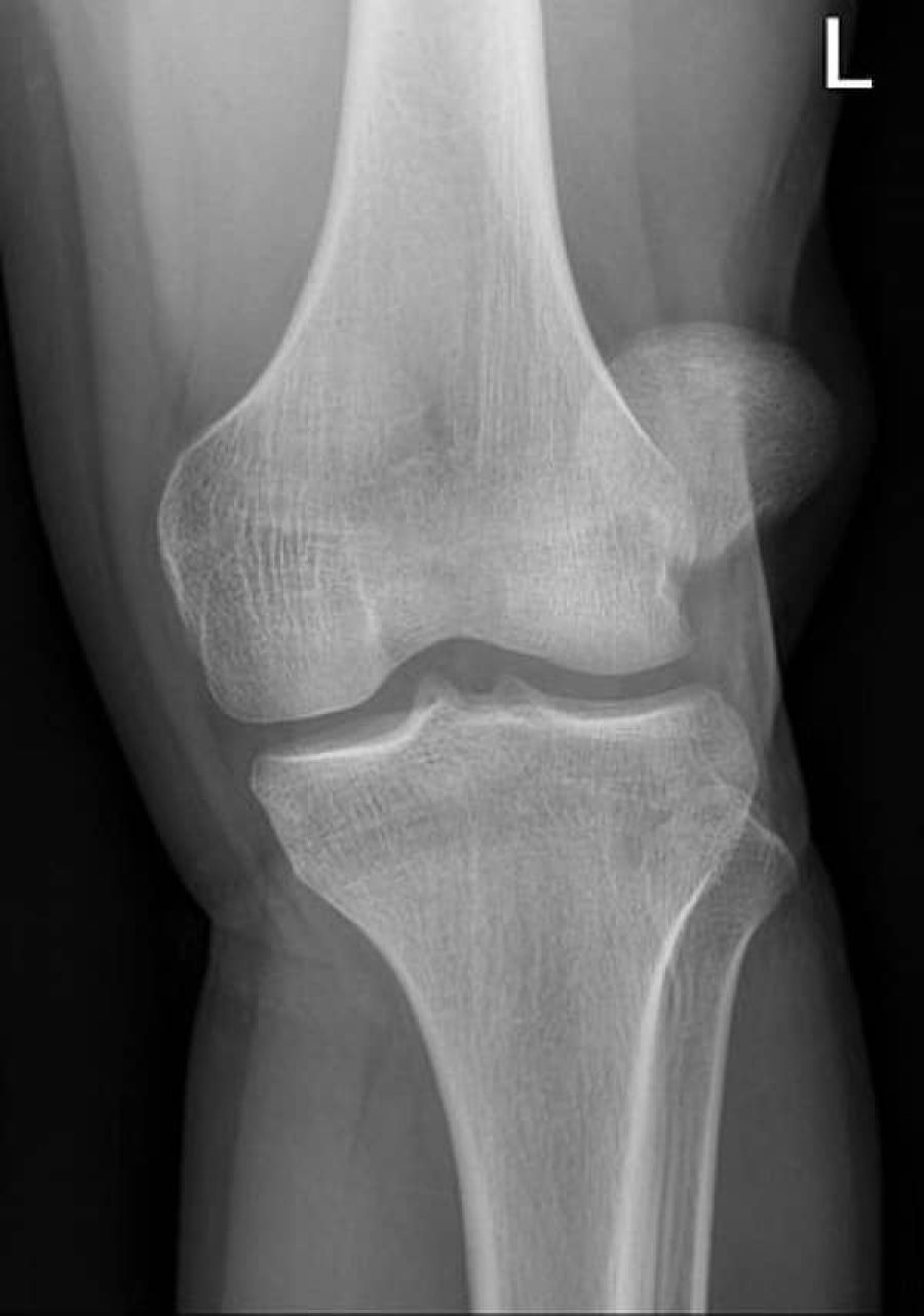
- X-rays; To exclude associated fractures (osteochondral, avulsion); subluxation will be seen on a lateral view
- CT: To measure tuberosity tibia-trochlea groove distance
- MRI: To differentiate degree of tear; to rule out osteochondral fractures
- Indicated in young patients with primary dislocations
Outcome Measures
- Fulkerson functional scale
- Lysholm knee scale
- Knee injury and osteoarthritis outcome score
- Knee outcome survey
- Lower extrimity function scale
- McGill pain questionnaire
Medical Management
Conservative Management
Indication:
- Primary patellar dislocation
In cases where the patella was not relocated spontaneously, it can be done under regional anaesthesia. Conservative management after reduction include: - Immobilization for 6 weeks (cylinder cast/back slab/knee range of motion brace)
- Medication:
- Supplements like glucosamine and
- NSAID’s
Conservative treatment is the most common treatment after primary patellar dislocation.
Surgical Management
Surgical management is done arthroscopically, with or without surgical repair of the torn retinaculum or immediate patellar realignment
Indications:
- Recurrent/chronic dislocation
- Patellofemoral symptoms
- Associated osteochondral fracture or major chondral injury
- Substantial disruption of the medial patellofemoral ligament)- vastus medialis obliquus-adductor mechanism
- Laterally subluxated patella on the plain Mercer-Merchant view with normal alignment on the contralateral knee
- Failed conservative management
Surgical stabilization significantly reduces the redislocation rate of primary traumatic patellar dislocation in the young adult population, but is associated with a higher risk of patellofemoral joint osteoarthritis. Initial post-operative management consists of pain management, physiotherapy and cryotherapy .
Types of Surgery
- Lateral release: Release of tight lateral retinaculum to allow more medial tracking of the patella.
- Indication: Mild patellar instability
- Medial patellofemoral ligament reconstruction / proximal realignment
- Balance the patellar tracking to more natural (medial) alignment
- Often done with a lateral release
- Indication: Severe patellar instability
- Distal realignment / anteromedialisation
- Transferring of the tibial tubercle (where the patellar tendon attaches to the tibia). The bony attachment of the tendon is moved more medially to allow the patella to track normally
- Used in conjunction with the lateral release and/or the medial patellofemoral ligament reconstruction.
- Indication: Severe patellar instability
Physiotherapy Management
Goals:
- Improve function
- Prevent further dislocation:
- Taping: Lateral reinforcement will reduce the movement of the patella (to prevent dislocation)
Physiotherapy modalities include: - Prevention of re-dislocation:
- Taping: Lateral reinforcement will reduce the movement of the patella (to prevent dislocation)
- Bracing
- Reassurance and behavioural modification
- Improve range of motion:
- Manual therapy knee
- Knee mobilisations
- Combination therapy
- Strengthening exercises:
- Quadriceps, hamstrings, adductors, hip and lower abdomen
- Closed kinetic chain exercises are recommended
- Stretching:
- Improve flexibility of hamstrings and quadriceps
- Proprioception: Improve stability of the knee